GLYCOLYSIS

Video

The Video gives a visual explanation of glycolysis which I think provides useful in learning this may seem like a long complicated process. Guys, it is not difficult. It is just a little long and may be a little tedious but what do we have to lose. This is my interpretation of the video as well as some of my own knowledge in point form.
(1) Glycolysis has two main steps broken down into 5 enzyme catalyzed steps. In the first step glucose is phosphorylated and is converted to two glyceraldehydes-3-phosphate molecules. It is referred to as the investment stage. Whereas the second stage of glycolysis is referred to as the payoff phase where there is oxidative conversions of glyceraldehyde-3-phosphate to pyruvate and coupled formation of ATP and NADH molecules.
(2) Investment Stage Broken Down:
STEP1: Glucose is converted to glucose-6phosphate, the first irreversible reaction using ATP which is converted to ADP. It is catalysed by hexokinase and it cofactor Mg2+.
STEP 2: Glocuse-6-phosphate is converted to its isomer fructiose-6-phosphate which is a reversible reaction catalysed by phosphohexose isomerase. Mg2+ is again a cofactor.
Step 3: Another reaction where ATP is used and converted to ADP. This is an irreversible reaction catalysed by phosphofructose kinase 2 (pfk2). This enzyme is the most important regulatory enzyme in glycolysis. Fructose-6-phosphate is converted to fructose-1,6-phosphate.
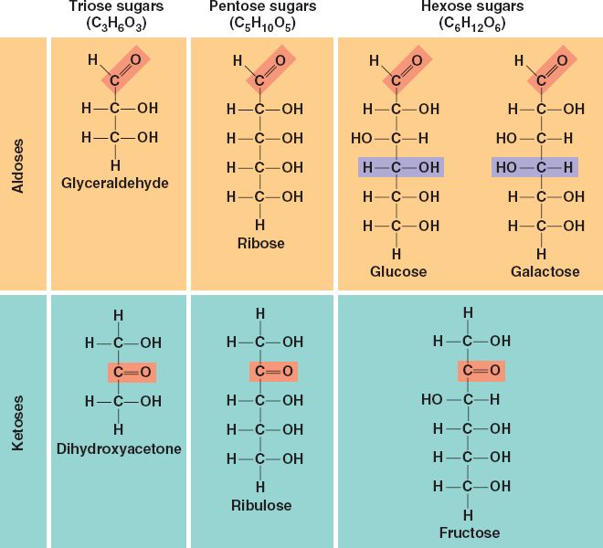
STEP 4: Fructose-1,6-phosphate is converted into two, three Carbon compounds; glyceraldehydes-3-phosphate and dihydroxyacetone phosphate. The enzyme that catalyses this reaction is aldolase which is a reversible reaction.
STEP 5: Dihydroxyacetone phosphate is converted to its isomer, glyceraldehydes-3-phosphate catalysed by triose phosphate isomerase. This is a reversible reaction.
NOW WE ENTER THE 2ND STAGE (PAY OFF)
STEP6: Each glyceraldehydes-3phosphate is converted to 1,3-bisphosphateglycerates. In this reaction an inorganic phosphorous source converts two NAD+ to NADH + H. This is a reversible reaction catalysed by glyceraldehyde-3phosphate isomerase.
STEP 7: This is a dephosphorylation reaction where two molecules of ADP is converted to two molecules of ADP. The 1,3 bisphosphoglycerate molecules are converted to 3-phosphoglycerate by phosphoglycerate kinase and it cofactor Mg2+. It is a reversible reaction.
STEP 8: the two molecules of 3-phosphoglycerate is converted to 2-phosphoglycerate which is a reversible reaction catalysed by phosphoglucerate mutase.
STEP 9: The two 2-phosphoglycerate is converted to phosphoenolpyruvate catalysed by enolase. This is a reversible reaction in which two molecules of H2O is released.
STEP 10: the two molecules of Phosphoenolpyruvate is converted to two molecules of pyruvate. In this reaction two molecules of ADP is converted to ATP. Pyruvate kinase catalyses this reaction which is irreversible.

ENZYMES AND INHIBITION

Aside

Enzymes are biological molecules which catalyze metabolic reactions necessary to drive all life cycles. The do this lowering the activation energy of the reaction. However they do not change equilibrium, only the rate at which equilibrium is reached. Thus they do not change the free energies of the reactant or the products. Activation energy is the minimum amount of energy for a reaction to occur which is lowered by enzymes. This is the point of the reaction where the arrangement of intermediate structures between reactant and product is at its highest.

All enzymes are specific, some more than others. When an enzyme only reacts with a one certain substrate, it is said to have an absolute specificity. On the other hand some enzymes act on substrates of a particular functional group or side chains, having relative/group specificity and others, the least specific act on certain bonds. That is linkage specificity. Also there are stereochemical specific enzymes which act on a specific isomer.

Enzymes speed up the rate of reaction and inhibitors deter them from doing this. There are four types of irreversible inhibitors; competitive, non competitive, uncompetitive and mixed competitive inhibition. Inhibitors are important so to ensure that a surplus of product is regulated depending on the requirements of the body. 

Image

Competitive inhibitors compete for the active site with substrates where they bind. Competitive inhibitors can do this since they resemble the substrates. This will decrease the affinity of the active site to the substrate to an extent depending on how much inhibitor to substrate concentration in present. The higher the substrate concentration the inhibitor effect will decrease. Thus the Km increases and Vmax remains the same as show on graph (a)

Noncompetitive inhibitors bind on any site that is not the active site. It makes the enzyme undergo a conformational change. Competitive inhibitor can bind to either the free enzyme or the enzyme substrate complex. It does not resemble the substrate. The km remains the same and the Vmax decreases as shown in graph (b). Thus increasing enzyme substrate will not prevent the effect of the inhibitor. Mixed inhibitors have similar charactoistics however they differ by their kinetics in that they either decrease or increase km values and

Uncompetitive inhibitors bind only to the enzyme-substrate complex, not on the active site. Therefore there is no need for this inhibitor to resemble the substrate. Look at graph (c) and you will notice that the lines representing enzyme catalyzed reaction and inhibited enzyme catalyzed reactions you will notice the lines are parallel and you can see that the km is reduced by the amount that Vmax is being reduced.

Phenolase, peroxidase and xanthine dehydrogenase are enzymes, all of which catalyse oxidation reactions and therefore belong to the first class of enzymes, oxidoreductases. Phenolase also call polyphenol oxidase has the enzyme commission number 1.10.3.1.  Copper (II) ions is its inorganic cofactor.

AMINO ACIDS AND PROTEINS

Image


All amino acids have this basic structure. They differ by their R groups. The smallest R group is a hydrogen atom. The R groups can be of varying lengths of C chains whether they are polar, uncharged R groups, non polar aliphatic R groups, aromatic R groups or positively charged R and negatively charged R groups. Hence the R group is used to classify R groups.

Amino acids undergo oxidation reactions with each other to form peptide bonds between each other. Many amino acids can be linked by peptide bonds to form proteins. Proteins make most of the enzymes that allow the catalysis of metabolic reactions necessary for life to continue.


MCQS

Aside

HOW ABOUT A NICE SHORT AND SWEET QUESTIONAIRE FOR YOU

Select the correct multiple answer using one of the keys A, B, C, D OR E as follows:

  1. A. 1,2 and 3 are correct
  2. B. 1 and 3 are correct
  3. C. 2 and 4 are correct
  4. D. Only 4 is correct
  5. E. All are correct

I. In what type of cell would mitochondria be found in relatively large quantities?

1) Liver cells

2) Nerve cells

3) Muscle cells

4) Euthrocytes

II. In what type of cell would lysosomes be found in the greatest quantities?

1) Eukaryotes

2) Muscle cells

3) Nerve cells

4) Liver cells

III. Which statement/s best describes the golgi-apparatus

1) The golgi-apparatus receives mRNA which it uses to transcribe for proteins and then modifying the proteins and or adding signal sequences befoor it shipped off.

2) The golgi-apparatus receives vesicles containing proteins on it trans side which will then be modified, signal sequence may be added and packaged to be shipped off in vesicles on the cis side.

3) The golgi-apparatus is the site of lipid synthesis.

4) The golgi-apparatus receives vesicles containing proteins on it cis side which will then be modified, signal sequence may be added and packaged to be shipped off in vesicles on the trans side.

IV. Mannose and glucose are epimers. This means that the orientation of a H atom and an OH group differ only on a certain C atom. Which C atom does this occur?

1) 1

2) 2

3) 3

4) 4

V. Some importance functions of carbohydrates include:

1) Structure

2) Energy source

3) Precursor molecule

4) growth

Need help studying carbohydrates

Click to access Chapter_17.pdf

Hey guys so this pdf was really helpfull.. Maybe it will be for you to. It is all abou the carbohydrates
It includes a few things i didnt tlk about in my posts including some starch and cellulose. Those are important to know. Also a small bit on lactose.
Also another important thing to remember some functions of carbohydrates:
1) energy source: we need to break down carbohydrates to obtain its stored energy to drive everyday metabolic reactions.
2) storage: listen we cant let sucrose build up in our blood. Yes it gives us energy but guys remember too much of a good thing is a bad thing. We animals use glycogen and plants use starch for storage. In animals hormones such as insulin and glucagon regulate concentrations of sucrose in our blood. Insulin convert glucose to glycogen. Glucagon convert glycogen to glucose.
3) structure: the more complex carbohydrates provide good, strong and rigid cell walls; cellulose for plant and murein for prokaryotes and insects have exoskelotons made of chitin.

Yup yup yup

20130412-052300.jpg

CARBOHYDRATES

Aside

CHARACTORISING CARBOHYDRATES
MONOSACCHARIDES DISACCHARIDES OLISACCHARIDES POLISACCHARIDES
One carbohydrate sub-unit. The simplest of the carbohydrateCan be further classified based on the length of the base carbon chain:

Triose – 3 C atoms

Tetros – 4 C atoms

Pentose – 5 C atoms

Hexose – 6 C atoms

Heptose – 7 C atoms

Can also be further classified based on the presence of either an aldyhyde or ketose fubctional group.  

Carbohydrate consists of two monosaccharides attached by a glycosidic bond. Examples:Sucrose – glucose and fructose

Lactose – glucose and galactose

Maltose – glucose and glucose


Carbohydrate consists of 5 to 10 monosaccharides attached by a glycosidic bond. Long chain carbohydrates consisting of hundreds or thousands of monosaccharides joined by glycosidic bonds.Examples: Amylose

Amylopectin

Cellulose


ALDOSE FUNCTIONAL GROUP vs KETOSE FUNCTIONAL GROUP

05_03Monosaccharides

ANOMER ISOMERISM

A monosaccharide can carry an alpha configuration or a beta comfiguration

Let us look at glucose: What is the difference between alpha (α) and beta (β) glucose?

image-3

Both pictures represent an isomer of glucose but take a closer look and tell me if you find a difference between the two… and guys it’s not a trick question. Yes they are both glucose and yes there is a difference.

Well look at the highlighted OH groups on both molecules; one is above the plane of the anomeric C atom and the other is below. So on the α glucose molecule the OH group is below the anomeric carbon and on the β glucose molecule the OH group is above the anomeric C atom. BUY OOOOOOO! What is that new word I’m using ‘anomeric’ what is that? Well that simply means the carbon atom that now the stereocenter as a result of cylization. It allows for this anomer isomerism.

CHIRALITY: simply means that there are four different bond attached to the central carbon atom. Molecules that express chirality are superimposable and have its mirror image.

D vs L ISOMER

glucos

The image on the right shows two isomers of glucose in the fischer projection; D-isomer and L-isomer.

NB: the functional group in glucose is the aldehyde group.

The D isomer shows that the OH group attached to the chiral carbon furthest from the functional group is on the right.

The L-isomer shows the OH group attacheched to the chiral carbon furthest from the functional group is on the left. 

images (32)

EPIMERS

Yet another type of isomerism. Epimers only differ in atomic configuration around one C atom. Take the example seen on the right ; mannose and glucose are epimers of each other since they differ only by the orientation of the OH group and H atom on C2. Likewise, galactose differs from glucose similarly only on C4.

GLYCOSIDIC BOND

glycosidic-linkage

Looking at the diagram on the right we can see a glycosidic bond being formed in a process called condensation.

2 α glucose molecules are bonding to form the maltose. The O atom on C1 of the 1st molecule is lost and the OH group. on C4 of the 2nd molecule is lost i.e. a water molecule is lost. The oxygen on C1 of the1st molecule can then bond with the C4 on the 2nd molecule. The reverse reaction would be hydrolysis.

Now do you think you can name some carbohydrates based on what we have learnt today?

maltose

If these two α glucose molecules under condensation what will we call the new product.

Well, the bond will take place between a C1 and a C4 and we know they are both α molecules and two glucose molecules make maltose

α1-4 maltose.

How about this one?

images (33)Since the answer is already there I’ll let you tackle this one on your own.

Last one. Remember guys practice makes perfect!! 

And this time I’m not giving you the answer

images (34)


JUST A THOUGHT

So just a thought…..Well cells are the most basic unit that all living specimen are composed of right! Well atoms are the most basic structure that all matter is composed of. The cell has organelles which are vital to its function and atoms have sub atomic particle that are vital to their function. Basically what i am gettin at is simply that cells are to living specimen what atoms are to animate to matter

20130402-023234.jpg

Endosymbiosis Theory

Video

ENDOSYMBIOSIS THEORY: – The video explains the following
• Origin and explanation of the meaning of endosymbiosis – meaning things live together on the inside
• Using simplified representation of cells he shows cells that could not produce energy nor could they produce food (photosynthesize).
• There were cells that could produce energy and cells that could produce food which were engulfed by those that could.
• A symbiotic relationship was then established. The cells that could respire and photosynthesize were housed and the cell that engulfed the others now had means of respiring aerobically and producing its own food. That is endosymbiosis.
• Proof to this theory is evident due to the similar structure of DNA molecules found within the eukaryotes mitochondria and cytoplasm compared with that found in prokaryotes.
The video is very vague and only gives a simplified image or idea of the symbiotic theory. Also it does not provide all the evidence which supports the theory. Some information that the video lacked is as follows:-
• Primary endosymbiosis is the case where a larger eukaryotic cell engulfs a prokaryotic cell. Take for example if the eukaryotic cell engulfs an aerobic prokaryote, the eukaryote becomes aerobic.
• Secondary endosymbiosis is the case where a eukaryotic cell engulfs another eukaryotic cell that has already undergone endosybiosis.
• When a cyanobacterium is engulfed, eukaryotic cells become photosynthetic.
Evidence: – Mitochondria and chloroplasts have similar traits to that of prokaryotic bacteria.
• Both mitochondria and chloroplasts have double membranes. The outer membrane resembles that of the eukaryote but the inner membrane resembles that of the prokaryotic cell.
• Both mitochondria and chloroplasts have their own single circular chromosome.
• Both mitochondria and chloroplast have 70s ribosomes.
• Both mitochondria and chloroplasts reproduce via binary fission.
• Both mitochondria and chloroplast are often very similar in size; 1 to10 microns.
• Electron transport chains are present in both mitochondria and chloroplasts.

Endosymbiotic_Theory_by_RiceandBeans

CELLS

Why hello my fellow biochem blogarians and other readers. Well let me say this before we get to business. We have about five weeks until exams, continuous incourse quizzes and several assignments. Two of these assignments are literature reviews, my worst nightmare. So usually I would complain and complain to myself.  My thoughts would be full of ideas on how to doom my lecturers and what not. Jk jk… I would totally never say that on this blog if it were true. No but it is long past time I started finishing these assignments so today that is my goal. Procrastination has been for far too long my greatest crime. So anyway lets tlk a little bit about the cell. Most of this you may however already know and this piece will serve as a form of revision I like to call regurgitation.

So to business                                                                                                                                                                  Our topic for today is cells and when we think of cells, we think of tiny entities. Actually today we know that they are the basic fundamental units of all life. Hence, the term unicellular and multi-cellular is used to describe all organisms. Mathhias Scheilden is a German botanist who realized that plants were made up of cells. Then Theodor Schwann realized that animals’ most basic composition is on a cellular level. Rudolf Virchow whom was actually a physician discovered that cells could only come from other cells while studying viruses and the first man to ever see a cell was Robert Hooke.  He saw a cork cell under a much less advanced microscope than we see today while doing his research. These scientists of the seventeenth century contributed to what we know now as the cell theory that states that the most basic unit of structure and function of all life forms are cells and that living cells can only be reproduced by other living cells.

                              kingdoms

Two major types of cells: Prokaryotes and Eukaryotes

Both cell types are composed of individual structures which collectively allow the functioning of the cell.

There are structures similar to each as well as unique to both.

                                       prokaryotic_cell_h650px

PROKARYOTIC CELLS: The defining feature of prokaryotes is their lack of a nucleus.

CELL WALL – A layer that surrounds the plasma membrane. Composed of peptidoglycan molecules cross linked with each other (mostly murein). This structure gives it its rigidity. The function of the cell wall is to provide support and maintain hydrostatic pressure. 

CELL MEMBRANE/PLASMA MEMBRANE –  A layer whose limits are within the cell wall and the cytoplasm.  It is composed of a bilayer of phosphate heads and lipid tails, embedded in which are transport proteins.  Thus the plasma membrane is semi permeable. The function is to control what passes through the plasma membrane.

INFOLDING OF PLASMA MEMBRANE – Compensates for the lack of membrane bound organelles. That is proteins needed for metabolic pathways such as respiration and photosynthesis are made here.

GINETIC MATERIAL– DNA held on circular chromosome like structures which have no associated histone proteins. found inthe nucleiod.

PLASMIDS – A circular DNA molecule which reproduces independent to the rest of the cell.

CYTOPLASM – This is basically the contents of the cell. It is composed of all the organelles and a fluid filled cavity called cytosol.

RIBOSOMES – 70s ribosomes present in prokaryotes. RNA – protein complexes. Sites of protein synthesis.

PILLI – A sticky outer layer which allows for adhesion. It provides a mechanism for attachment to other cells during sexual reproduction.

CAPSULE – It surrounds the pilli and provides protection.

MESOSOME – Specific enfolding of the cell membrane which plays a role in cell replication/reproduction and or excretion of exoenzymes.  

FLAGELLUM – A tail like structure whose function is locomotion.  It moves with a whip like motion propelling the organism.                                                                     


EUKARYOTIC CELLS: The defining feature of eukaryotes is the presence of a nucleus.

                                  Animal_Cell_Diagram                                                                                Plant_Cell_Diagram


NUCLEOLUS – The nucleolus is found within the nucleus. Its function is to transcribe and assemble the ribosomes.

NUCLEUS – Considered the control center of the cell since it houses all genetic material that codes for all functional enzymes of the cell’s metabolic processes. It is surrounded by a double membrane which is porous.

ENDOPLASMIC RETICULUM – The nucleus outer membrane forms a continuous folding which forms the endoplasmic reticulum. Synthesized biochemical molecules are then shipped off in vesicles to the golgi apparatus.  

ROUGH ER – Where the endoplasmic reticulum is studded with ribosomes, looking like sheet s of bumpy ER. Here proteins are synthesized.

SMOOTH ER – This endoplasmic reticulum lacks ribosomes and looks like smooth tubular membrane. It is a storage organelle which holds ions that may be needed at a later time. It is also the site of steroid synthesis.

GOLGI APPARATUS – Here vesicles that pinch off from the endoplasmic reticulum fuse to the side of the goglgi apparatus closer to the nucleus. This side is referred to the cis side. In this structure the different biomolecules are separated, modified and signal systems are attached and then packaged in vesicles. These pinch off the Trans side. This is the side farthest from the nucleus.  The vesicles move to their designated destinations and carry out their roles.

LYSOSOMES – Vesicles that pinch off the trans side of the golgi apparatus. It contains lipases, nucleases, and proteases. These vesicles fuse with other vesicles and their enzymes break down lipids, protein and nucleic acids respectively. The vesicles that they have fused with would have been engulfed thus lysosomes deal with exogenous degradation.

PROTEASOMES – These are similar to lysosomes, however their enzymes only break down proteins made within the cell. That is endogenous proteins.

PEROXIOSOMES -These are similar to lysosomes and proteasomes. They contain oxidative enzymes which break down amino acids and fatty acids

CENTRIOLES – A cylindrical structure involved in the mitotic spindle formation and in the completion of cytokinesis.

CHLOROPLAST – Site of light energy capture for conversion to chemical energy for a food source through a process called photosynthesis.

CELL WALL – Similar function to that of the cell wall in prokaryotes however composed of a different material in plants; cellulose, a complex polysaccharide. The cell wall in fungi also has the same function. However it is made of another polysaccharide known and chitin.

There are organelles and structure that are common to both animal and plant cells. There are other organelles and structures that are unique to animal cells and plant cells.

Some organelles and structures that is unique to animals: lysosomes, proteasomes, peroxisomes, centrioles.

Some organelles that are unique to plants: cell wall, chloroplasts

So two more points I would like to make: Firstly remember we said that we think of cells as small entities. Well do you still think so knowing that they are composed of so many different structures?

Let me answer you: OF COURSE, you see cells have to be tiny because they require getting rid of waste products as well as obtaining nutrients. The way they do this is by diffusion. If the cell gets too big then the diffusion rate will not sustain cellular life.

Then you might say that cell can be really really small but they cannot be too small guys. They need to be just big enough to hold all the DNA and other structures important to sustain the cell’s life. 

smiley-face-grin

SOURCES:

http://www.ric.edu/faculty/ptiskus/six_kingdoms/

http://askabiologist.asu.edu/plasmids

http://www.enchantedlearning.com/subjects/plants/cell/

http://www.biologyjunction.com/cell_functions.htm

http://www.4to40.com/science/print.asp?p=Parts_of_a_Cell&k=Eukaryotic_Cell